Embryonal, non-rhabdoid CNS tumours (former CNS-PNET) and pineoblastoma – Brief information
This text provides information on embryonal, non-rhabdoid tumours of the central nervous system and on pineoblastoma, a tumour of the pineal gland. The reader will be informed about the characteristics and subtypes of these diseases, their frequencies, causes, symptoms, diagnosis, treatment, and prognosis.
Author: Maria Yiallouros, Reviewer: Dr. med. Martin Mynarek, English Translation: Dr. med. Gesche Riabowol (nee Tallen), Last modification: 2024/03/29 https://kinderkrebsinfo.de/doi/e199450
Table of contents
General information on the disease
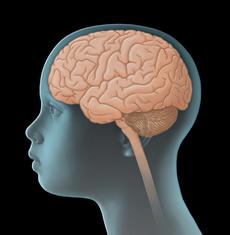
Embryonal, non-rhabdoid tumours of the central nervous system (until recently known as primitive neuroectodermal tumours of the central nervous system, CNS-PNET) and pineoblastoma are solid tumours that arise from brain tissue or spinal cord tissue. Since they directly originate from the CNS, they are also called primary CNS tumours, thereby differentiating them from malignant tumours of other organs that have spread (metastasised) to the CNS.
Both, embryonal CNS tumours and pineoblastoma originate from extremely immature (undifferentiated) cells of the central nervous system, which divide at a high rate. Therefore, these tumours grow very fast. They look very alike under the microscope and also share various features with medulloblastoma, an embryonal tumour of the cerebellum.
Embryonal, non-rhabdoid CNS tumours and pineoblastoma differ, for example, with regard to their location in the CNS: non-rhabdoid CNS tumours usually grow in the upper part of the brain (supratentorially), mostly in the cerebral hemispheres [see cerebrum]; hence they were previously called supratentorial PNET (stPNET) in order to differentiate them from the also embryonal medulloblastoma. Very rarely, they develop in other areas of the CNS. Pineoblastoma usually grows in the area of the pineal gland, a small endocrine organ in the centre of the brain (pineal region).
Both tumour types show an aggressive growth pattern. Embryonal non-rhabdoid CNS tumours often spread from one cerebral hemisphere into the other and/or into the meninges, from where they invade additional CNS tissue. Pineoblastoma may spread from the area of the pineal gland into other regions of the brain and spine as well. Metastasis outside the CNS, for instance to bones, bone marrow, lung, or lymph nodes, is rare.
Tumour types
The group of embryonal, non-rhabdoid CNS tumours (prior to the update of the WHO classification known as CNS-PNET) includes different tumour types, which differ regarding their histological and, partly, also to their molecular features.
According to the current classification of the World Health Organization (WHO) for tumours of the central nervous system (WHO classification 2016), the tumours considered here include, above all, the “embryonal tumours with multilayered rosettes (ETMR)”, which – depending on harbouring or not harbouring a certain genetic alteration on chromosome 19 (the so-called C19MC amplification) – are called either ETMR C19MC-altered or ETMR NOS (short for: not otherwise specified). Additional tumours of this group are, for example, the rare medulloepithelioma and other embryonal, non-rhabdoid CNS tumours that have not been otherwise specified.
Due to their rarity and similarity regarding the course of the disease, many authors considered pineoblastomas and embryonal, non-rhabdoid CNS tumours as one group in the past. Today, however, it is well-known that a pineoblastoma differs substantially from other embryonal CNS tumours on the molecular level and, therefore, needs to be dealt with as an independent tumour type.
Embryonal, non-rhabdoid CNS tumours as well as pineoblastoma are defined as high-grade malignant tumours (WHO grade IV).
Incidence
Embryonal, non-rhabdoid CNS tumours and pineoblastoma are rare: they account for approximately 2 % of all CNS tumours in childhood and adolescence. In Germany, about ten children and adolescents under 15 years of age are newly diagnosed with one of these tumours each year. Embryonal CNS tumours are most frequent in children within their first years of life – the patients’ average age at diagnosis ranges between three and four years. Boys and girls are almost equally affected. Pineoblastoma is mostly diagnosed in children and young adults.
Causes
Embryonal, non-rhabdoid CNS tumours and pineoblastoma are caused by a malignant transformation of nerve tissue cells. The reasons for tumour development have not been completely found out yet. It is well-known, though, that radiation of the brain, for example as received by children with certain forms of leukaemia or with eye cancer (retinoblastoma), leads to an increased risk of developing a CNS tumour later in life.
In addition, it has been shown that embryonal CNS tumours and pineoblastoma are sometimes associated with certain genetic and chromosomal abnormalities in the tumour cells. The resulting impairments of cell development and cell communication may be contributing factors promoting the transformation of a healthy into a cancer cell. However, since embryonal, non-rhabdoid CNS tumours and pineoblastoma are rare, only a few molecular abnormalities that might be responsible for causing the disease have been identified yet.
Good to know: rarely, pineoblastoma can be associated with hereditary retinoblastoma and thus genetic alterations of the retinoblastoma gene (so-called trilateral retinoblastoma). More information on trilateral retinoblastoma can be found in our Retinoblastoma chapter.
Symptoms
Due to the uncontrolled and aggressive growth pattern of embryonal, non-rhabdoid CNS tumours and pineoblastoma, symptoms typically develop and deteriorate fast. The presenting symptoms of these tumours (like other tumours of the central nervous system) primarily depend on the patient’s age, the site and size of the tumour as well as its pattern of spread within the CNS. The following general (nonspecific) and local (specific) symptoms can occur:
General (nonspecific) symptoms
Unspecific general symptoms occur independently of the tumour’s location. They may be similar to and therefore mimic other, non-CNS diseases. General symptoms of a child or adolescent with a CNS tumour may include headaches and/or back pain, dizziness, loss of appetite, nausea and vomiting (particularly after getting up in the morning), weight loss, increasing fatigue, inability to concentrate, school problems, mood swings, and character changes as well as developmental delay, to name a few.
Major reason for these symptoms is the slowly but continuously increasing intracranial pressure (ICP). An elevated intracranial pressure may be caused by the growing, thus more and more space-occupying tumour within the bony skull. It may as well be due to the tumour blocking the regular flow of the cerebrospinal fluid, thereby forming hydrocephalus. In babies or small children with soft spots (open fontanelles), elevated intracranial pressure and hydrocephalus typically present with a bulging fontanelle or a larger than expected head circumference (macrocephalus), respectively.
Local (specific) symptoms
Local symptoms may indicate the tumour location and, thus, which functional regions of the CNS might be affected. For example, a tumour in the hemispheres of the cerebrum or in the diencephalon can be associated with seizures and/or motor deficits. Also, vision impairments, speech disorders, behavioural and sleep problems, as well as moodiness and altered appetite regulation may, although to a lesser extent, be indicative of tumour location. Pineoblastoma can cause trouble with eye movements, in particular limitations of upgaze. This vision impairment results from the tumour’s specific location in the diencephalon and is also known as Parinaud syndrome.
Good to know: Not all patients presenting with one or more of the symptoms mentioned above do have an embryonal CNS tumour, a pineoblastoma or another type of brain tumour. Many of these symptoms may also occur with other, harmless diseases that are not associated with a brain tumour at all. However, if certain symptoms persist or get worse (for example repetitive headaches or rapid increase of head circumference in a young child), a doctor should be seen to find the underlying reason. In case it is a brain tu-mour, treatment should be started as soon as possible.
Diagnosis
If the paediatrician thinks that the young patient’s history (anamnesis) and physical examination are suspicious of a tumour of the central nervous system (CNS), the child should immediately be referred to a hospital with a childhood cancer program (paediatric oncology unit), where further diagnostics can be initiated and performed by childhood cancer professionals. Very close collaboration between various specialists (such as paediatric oncologists, paediatric neurosurgeons, paediatric radiologists, to name a few) is required, both to find out, whether the patient really suffers from a malignant CNS tumour and, if so, to determine the tumour type and the extension of the disease. Knowing these details is absolutely essential for optimal treatment and prognosis.
Tests to secure diagnosis
The initial diagnostic procedures for a young patient presenting with a suspected CNS tumour at a childhood cancer centre include another assessment of the patient’s history, a thorough physical/neurological exam and imaging diagnostics, such as magnetic resonance imaging (MRI) or (less often) computed tomography (CT). These tests help find out exactly whether the patient has a tumour of the central nervous system. Also, localisation and extent of the tumor, its demarcation regarding adjacent tissue as well as a potential hydrocephalus can be diagnosed by these imaging techniques very well.
In order to validate the final diagnosis, histological and molecular analysis of surgically obtained tumour tissue (biopsy) is required. Usually, this is done using the tissue obtained during surgical tumour removal. The extent of histological and, especially, molecular genetic workup has been substantially increased over the past years. Today’s option of using modern lab techniques makes it possible to identify molecular tissue characteristics that do not only help finalize the diagnosis, but can also provide information on what to expect regarding the course of the disease (such as growth behaviour). Therefore, molecular diagnostics will play a major role in designing future treatment strategies.
Tests to assess spread of disease
Once the diagnosis of an embryonal CNS tumour or a pineoblastoma has been confirmed, additional tests are required to assess the extent of the disease within the central nervous system (CNS). Apart from MRI scans of the complete CNS (brain and spine) for macroscopic metastases, these tests also include microscopic checking of the cerebrospinal fluid (CSF) for tumour cells in the spinal cord (which are not visible by MRI scan). Cerebrospinal fluid is mostly obtained from the spine in the lower back (lumbar puncture), since the risk of the puncture needle damaging the spinal cord is lowest at the lower back level.
Tests before treatment begins
In preparation for the intensive treatment of the brain tumour, further investigations are performed, such as electrocardiography (ECG) and echocardiography to check cardio function. Furthermore, additional blood tests are needed to assess the patient’s general health condition and to check whether the function of certain organs (such as liver and kidneys) is affected by the disease and whether there are any metabolic disorders to be considered prior or during therapy. Any changes occurring during the course of treatment can be assessed and managed better based on the results of those initial tests, which thus help to keep the risk of certain treatment-related side effects as low as possible.
Also, the patient’s blood group needs to be determined in case a blood transfusion is required during treatment. In sexually mature females (which means after they have experienced their first menstruation), a pregnancy test is recommended prior to treatment as well.
Good to know: Not every patient needs the complete check-up. On the other hand, tests might be added that haven't been mentioned here, depending on the individual situation of the patient. Your caregivers will inform you and your child, which diagnostic procedures are individually required in your child’s situation and why.
Treatment planning
Once the diagnosis and extent of a CNS tumour has been confirmed, treat-ment planning starts. In order to provide the patient with the best possible individual and risk-adapted therapy, the treatment team considers specific factors that are known to have an impact on the prognosis (so-called prognostic factors).
Important prognostic factors in case of an embryonal, non-rhabdoid CNS tumour or pineoblastoma are the type (subtype), localization, extent and spread of the tumour. Also, the biological (molecular) features of a tumour increasingly impact the choice of optimal treatment. In addition, the patient’s age and overall physical condition play an important role. Age at diagnosis, above all, determines whether the patient may receive radiotherapy or not. All these factors are included in treatment planning in order to achieve the best outcome possible for each patient.
Treatment
Treatment of children and adolescents with an embryonal, non-rhabdoid CNS tumour or with pineoblastoma should take place in a children's hospital with a paediatric oncology program. Only in such a childhood cancer centre, highly experienced and qualified staff (doctors, nurses and many more) is guaranteed, since they are specialized and focus on the diagnostics and treatment of children and teenagers with cancer according to the most advanced treatment concepts. The doctors in these centres collaborate closely with each other. Together, they treat their patients according to treatment plans (protocols) that are continuously optimised. The goal of the treatment is to achieve higher cure rates while avoiding side effects as much as possible.
Current treatment concepts involve neurosurgical tumour removal, chemotherapy and, depending on the patient’s age, radiotherapy.
Surgery
The first step in treating an embryonal CNS tumour or a pineoblastoma is surgery. Goal of surgery is gross (microsurgical) total tumour removal. This means that at the end of the surgical procedure, no tumour tissue can be identified through the surgical microscope. Frequently, however, total tumour resection cannot be achieved due to the localization of these tumours.
In most patients, neurosurgical intervention results in normalising the flow of cerebrospinal fluid (CSF). Patients initially presenting with hydrocephalus may need a transient hydrocephalus drainage prior to tumour removal or, sometimes, even a permanent drainage system later.
Additional, non-surgical treatment
Since embryonal CNS tumours and pineoblastomas tend to infiltrate adjacent tissue and, furthermore, often spread into other parts of the central nervous system via the cerebrovascular fluid (CSF), treating the tumour locally only is not sufficient. Therefore, surgery is followed by additional non-surgical treatment, comprising radiation therapy and/or chemotherapy.
Chemotherapy uses drugs (so-called cytostatic agents or cytostatics) that can kill fast-dividing cells, such as cancer cells, or inhibit their growth, respectively. Radiotherapy is done using energy-rich, electromagnetic radiation, given through the skin to the tumour region. Radiation causes DNA damage in tumour cells, thereby leading to cell death. Aside from this so-called conventional radiotherapy, particle-radiation with protons (also known as proton therapy) can be an option for some patients as well. This type of radiotherapy provides the benefits of better targeting the tumour area, thus sparing more adjacent, healthy tissue from the effects of radiation. Proton therapy is gaining an increasing importance in the treatment of children and teenagers with solid tumours.
Decision upon which therapy is to be applied (treatment modalities, intensity of chemo-/radiotherapy) is based on the patient’s age, the histological and molecular subtype of the tumour, certain biological risk factors, as well as on the extent of both metastases and surgical tumour removal.
Treatment options for pineoblastoma patients
After maximal possible tumour removal, patients with non-metastasised pineoblastoma, who are older than four years of age, will receive radiation of the complete central nervous system (craniospinal radiotherapy), followed by an additional boost to the tumour site. Radiotherapy is followed by a so-called maintenance chemotherapy, which includes multiple cytostatic agents. In cases of metastasis, treatment will be intensified, for example by giving higher doses of radiotherapy combined with a preceding chemotherapy (induction chemotherapy).
In children under four years of age, whose brain development is not completed yet, radiotherapy should be avoided or delayed in order to minimize the risk of serious late effects. Instead of radiotherapy after surgery, patients will receive chemotherapy with multiple agents. In some patients, radiotherapy may be an option later on. Some patients are also eligible for high-dose chemotherapy followed by autologous stem cell transplantation to increase the chances of survival.
Treatment options for patients with other embryonal CNS tumours
For other embryonal tumours of the central nervous system, various subtypes have been identified according to recent reports. These require individual treatment approaches depending on the presenting subtype.
Good to know: the details of how an individual patient is treated will be discussed between the responsible physician, the patient and the family.
Therapy optimising trials and registries
The majority of the children and adolescents with embryonal non-rhabdoid CNS tumour or pineoblastoma or a recurrence of these diseases receive therapy according to the treatment plans of therapy optimising trials or registries. Therapy optimising trials are standardised and controlled clinical trials that aim at steadily developing and improving treatment concepts for sick patients based on the current scientific knowledge.
Patients who cannot participate in any study, for example because none is available or open for them at that time or since they do not meet the required inclusion criteria, respectively, are often included in a so-called registry. One goal of a registry is to collect treatment data for research questions. Furthermore, the registry centre supports the doctors at site with (non-commital) treatment recommendations based on the most recent data on best treatment options, in order to provide the patient with optimal therapy even without the framework of a clinical study.
In Germany, a long-term therapy optimising trial (trial “HIT 2000”) for the treatment of children and adolescents with embryonal, non-rhabdoid CNS tumours (then named CNS-PNET) or pineoblastoma (also including patients with medulloblastoma and ependymoma) was closed in 2011. Many children’s cancer centres in Germany and Austria had participated in this trial. Currently, there is no open trial for patients with first diagnosis of embryonal CNS tumours or pineoblastoma. Newly diseased patients may, however, take part in the I-HIT-MED Registry (see below).
The following registries are available at the moment:
- I-HIT-MED Registry: Patients with embryonal, non-rhabdoid CNS tumours (before called CNS-PNET), who, for different reasons, cannot or do not want to participate in any currently available or open trial, can be enrolled in this registry (International HIT-MED Registry), regardless of the treatment given. These patients will receive treatment as per individually designed treatment plans. The goal of the registry is not to assess the feasibility of an ongoing trial, safety or efficacy of a certain treatment. It rather aims at collecting individual patient data for future analysis. The headquarters of the registry are located in the Children’s Cancer Centre at the University of Hamburg, Germany. The head of the study is Prof. Dr. med. Stefan Rutkowski.
- HIT-REZ Registry: Patients, whose disease does not respond to current treatments or with recurrent disease (relapse), respectively, can be enrolled in this registry, which has been open since January 2015. This registry does not serve to test new treatment regimens or drugs. However, the experts running the registry are providing treatment recommendations based on the most recent results obtained from national trials (for example from the HIT-REZ 2005 trial, which was closed in 2016) as well as international relapse trials. The headquarters of the registry are located in the Children’s Cancer Centre at the University of Essen, Germany. The head of the study is Prof. Dr. med. Gudrun Fleischhack.
Prognosis
The cure rates for children and adolescents with an embryonal, non-rhabdoid CNS tumour is, according to the German Childhood Cancer Registry, about 60% (5-year-survival rate). Overall prognosis for pineoblastoma patients is a bit more favorable.
However, in individual patience, prognosis is dependent on various factors. Since embryonal CNS tumours are a very heterogeneous group, the probability of survival can be different depending on the tumour type. Also, this stage of the disease, and the patient’s age play a significant role. Hence, children and adolescents with metastasized disease have generally a more unfavourable prognosis than patients with localized disease. Also, very young patients, for whom radiation therapy is not an option, have a rather unfavourable prognosis; only 20 to 30% of the patients experience long-term disease-free survival.
Note: The survival rates mentioned in the text above are statistical values. Therefore, they only provide information on the total cohort of patients with childhood Medulloblastoma. They do not predict individual outcomes.
In the context of cancer, the term „cure“ should rather be referred to as „free of cancer“, for even if current treatment regimens may help remove the tumour, the the tumour’s growth may have caused irrepairable damage to the brain or the treatment may be associated with late effects. Early detection and appropriate management of these long-term secondary effects typically require intensive rehabilitation and thorough long-term follow-up care.
 PDF Information on embryonal, non-rhabdoid CNS tumours and pineoblastoma (253KB)
01/09/2021, glossary added 29/03/2024
Literature 
- Kaatsch P, Grabow D, Spix C: German Childhood Cancer Registry - Anual Report 2018 (1980-2017). Institute of Medical Biostatistics, Epidemiology and Informatics (IMBEI) at the University Medical Center of the Johannes Gutenberg University Mainz 2019 [URI: https://www.kinderkrebsregister.de/ dkkr/ ergebnisse/ jahresberichte/ jahresbericht-2018.html]
- Fleischhack G, Rutkowski S, Pfister SM, Pietsch T, Tippelt S, Warmuth-Metz M, Bison B, van Velthoven-Wurster V, Messing-Jünger M, Kortmann RD, Timmermann B, Slavc I, Witt O, Gnekow A, Hernáiz Driever P, Kramm C, Benesch M, Frühwald MC, Hasselblatt M, Müller HL, Sörensen N, Kordes U, Calaminus G: ZNS-Tumoren. in: Niemeyer C, Eggert A (Hrsg.): Pädiatrische Hämatologie und Onkologie. Springer-Verlag GmbH Deutschland, 2. vollständig überarbeitete Auflage 2018, 359 [ISBN: 978-3-662-43685-1]
- Mynarek M, Pizer B, Dufour C, van Vuurden D, Garami M, Massimino M, Fangusaro J, Davidson T, Gil-da-Costa MJ, Sterba J, Benesch M, Gerber N, Juhnke BO, Kwiecien R, Pietsch T, Kool M, Clifford S, Ellison DW, Giangaspero F, Wesseling P, Gilles F, Gottardo N, Finlay JL, Rutkowski S, von Hoff K: Evaluation of age-dependent treatment strategies for children and young adults with pineoblastoma: analysis of pooled European Society for Paediatric Oncology (SIOP-E) and US Head Start data. Neuro-oncology 2017 Apr 1; 19: 576 [PMID: 28011926]
- Louis DN, Perry A, Reifenberger G, von Deimling A, Figarella-Branger D, Cavenee WK, Ohgaki H, Wiestler OD, Kleihues P, Ellison DW: The 2016 World Health Organization Classification of Tumors of the Central Nervous System: a summary. Acta neuropathologica 2016, 131: 803 [PMID: 27157931]
- Bode U, Zimmermann M, Moser O, Rutkowski S, Warmuth-Metz M, Pietsch T, Kortmann RD, Faldum A, Fleischhack G: Treatment of recurrent primitive neuroectodermal tumors (PNET) in children and adolescents with high-dose chemotherapy (HDC) and stem cell support: results of the HITREZ 97 multicentre trial. Journal of neuro-oncology 2014, 120: 635 [PMID: 25179451]
- Gerber NU, von Hoff K, Resch A, Ottensmeier H, Kwiecien R, Faldum A, Matuschek C, Hornung D, Bremer M, Benesch M, Pietsch T, Warmuth-Metz M, Kuehl J, Rutkowski S, Kortmann RD: Treatment of children with central nervous system primitive neuroectodermal tumors/pinealoblastomas in the prospective multicentric trial HIT 2000 using hyperfractionated radiation therapy followed by maintenance chemotherapy. International journal of radiation oncology, biology, physics 2014, 89: 863 [PMID: 24969797]
- Friedrich C, von Bueren AO, von Hoff K, Gerber NU, Ottensmeier H, Deinlein F, Benesch M, Kwiecien R, Pietsch T, Warmuth-Metz M, Faldum A, Kuehl J, Kortmann RD, Rutkowski S: Treatment of young children with CNS-primitive neuroectodermal tumors/pineoblastomas in the prospective multicenter trial HIT 2000 using different chemotherapy regimens and radiotherapy. Neuro-oncology 2013, 15: 224 [PMID: 23223339]
- Timmermann B, Kortmann RD, Kühl J, Rutkowski S, Meisner C, Pietsch T, Deinlein F, Urban C, Warmuth-Metz M, Bamberg M: Role of radiotherapy in supratentorial primitive neuroectodermal tumor in young children: results of the German HIT-SKK87 and HIT-SKK92 trials. Journal of clinical oncology 2006, 24: 1554 [PMID: 16575007]