High-Grade Glioma (HGG) – Brief Information
Highly malignant or high-grade gliomas (HGG) are tumours of the central nervous system (CNS). This text provides information about the characteristics and subtypes of the disease, its frequency, causes, symptoms, diagnosis, treatment, and prognosis.
Author: Maria Yiallouros, Editor: Maria Yiallouros, Reviewer: Prof. Dr. med. Christof Kramm, English Translation: Dr. med. Gesche Riabowol (nee Tallen), Last modification: 2026/04/10 https://dx.doi.org/10.1591/poh.hm-gliome.patinfo.kurz.1.20141217
Table of contents
General information on the disease
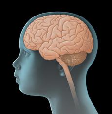
Highly malignant or high-grade gliomas (HGG) are tumours of the central nervous system (CNS). They are solid tumours arising from transformed cells of the brain and/or the spinal cord. Since they directly originate from the central nervous system, they are also called primary CNS tumours, thereby differentiating them from malignant tumours of other organs that have spread (metastasised) to the CNS.
High-grade gliomas in children and adolescents are rare. However, they show considerably malignant behaviour since they usually grow fast and frequently destroy healthy brain tissue. By being able to migrate within the central nervous system for various centimetres, high-grade gliomas can induce the development of new tumours. Without the appropriate therapy, high-grade gliomas can be lethal within only a few months. Due to their usually rapid and infiltrating growth, treatment is difficult.
Incidence
High-grade gliomas account for approximately 15–20 % of CNS tumours in children and adolescents. They appear in all age groups; yet, children aged younger than three years are rarely affected. Each year, about 70 children and adolescents younger than 15 years of age are newly diagnosed with a high-grade glioma in Germany. This corresponds to an incidence rate of 5 to 10 new diagnoses per 1.000.000 children per year. Boys are slightly more affected than girls.
Types of high-grade gliomas
Various types of high-grade gliomas (HGG) have been identified based on their location within the central nervous system (CNS), the cell of origin and the degree of malignancy.
High-grade gliomas of the brainstem
High-grade gliomas of the brainstem, such as “diffuse intrinsic pontine glioma" (as well as histologically and molecular genetically diagnosed "diffuse midline gliomas HR K27-alterated, CNS WHO-grade 4", as long as they are located in the brainstem), account for about one third of all HGG in children and teenagers. Due to their localization within the pons – an area of the brainstem, through which all important nerve tracts between extremities and brain are passing –, they can cause various severe neurological deficits and also result in a rapid and fatal course of the disease, even when they are small in size.
Note regarding (DIPG): Diffuse intrinsic pons glioma, which are usually diagnosed by imaging, frequently turn out as "diffuse midline gliomas histone 3 K27-alterated (CNS WHO-grade 4)" based on their histology and molecular genetic characteristics. According to the grading of the World Health Organization (WHO classification) of tumours of the central nervous system, these are classified as an entity of their own. However, despite their different terminology based on underlying diagnostics, DIPG and diffuse midline gliomas H3 K27-alterated often represent the same tumour type after all. Diffuse midline glioma H3 K27-alterated, however, do not only occur in the brain stem, but also in other parts of the brain (see below).
Gliomas in other parts of the central nervous system
About two thirds of high-grade gliomas are usually found in other parts of the CNS, especially in the cerebral cortex [cerebrum], but also in the cerebellum, diencephalon or spinal cord. These gliomas can be further subgrouped based on their histological and molecular characteristics, which provide inside into their origin and degree of malignancy. In general, these are tumours that are classified as CNS WHO-grade 3 or CNS WHO-grade 4 according to the current WHO classification (2021).
In addition, there are gliomas that are not classified according to the CNS WHO grading system. These include the gliomatosis cerebri, a type of tumour that (on the basis of imaging diagnostics) is typically characterised by a particularly extended infiltration and spread into adjacent tissue. Although its cells are frequently of low-grade malignancy, this tumour is classified as a highly malignant glioma because of its distinct growth pattern.
Good to know: The various types of high-grade gliomas differ in frequency and show different growth characteristics, different courses of the disease, different responses to treatment, and thus different outcomes (prognoses). This is also taken into account when planning the treatment (see chapter "Treatment planning").
Causes
High-grade gliomas originate from a malignant transformation of glial cells (glia). These are cells that, among other functions, provide support and protection for the brain’s nerve cells (neurons). Why glia cells become malignant in some individuals is not completely known yet.
It is known so far that children and adolescents with certain inherited diseases caused by genetic mutations (such as Li-Fraumeni syndrome or Turcot syndrome) have a higher risk of developing high-grade gliomas than their healthy peers. Due to the predisposition to tumours, such genetically determined conditions are also referred to as cancer predisposition syndromes.
Furthermore, certain genetic and/or chromosomal alterations are frequently observed in glioma cells. The resulting disruptions to further cell development and cell communication may play a causal role in the transformation of a healthy cell into a glioma cell. In general, such genetic alterations in tumour tissue are not inherited and most likely arise at a very early stage of development.
Also, radiotherapy of the brain (cranial irradiation), for example during treatment of leukaemia or retinoblastoma, a malignant tumour of the eye, increases the risk of developing a CNS tumour. Although not as frequently, chemotherapy can also promote the development of a high-grade glioma. However, for most children and adolescents no specific cause for the development of high-grade glioma has been identified so far.
Symptoms
Due to the tumours’ rapid growth, symptoms of children and adolescents with high-grade glioma usually develop within a few weeks or months. Similar to those of other tumours of the central nervous system (CNS), the presenting symptoms of high-grade gliomas primarily depend on the patient’s age, tumour site and size, and pattern of spread within the central nervous system. The following general (nonspecific) and local (location-related, specific) symptoms can occur.
General symptoms
General (unspecific) symptoms occur independently of the tumour’s location. They may be similar to and therefore mimic other, non-CNS diseases. General symptoms of a child or adolescent with a CNS tumour may include headaches and/or back pain, dizziness, loss of appetite, nausea and vomiting (particularly after getting up in the morning), weight loss, increasing fatigue, inability to concentrate, school problems, mood swings, and character changes as well as developmental delay, to name a few.
Major reason for these symptoms is the slowly but continuously increasing intracranial pressure (ICP). Elevated intracranial pressure may be caused by the growing, thus more and more space-occupying tumour within the bony skull (or spinal canal), but also by the tumour blocking the regular flow of the cerebrospinal fluid, thereby forming hydrocephalus. In babies or small children with soft spots (open fontanelles), elevated intracranial pressure and hydrocephalus typically present with a bulging fontanelle or a larger than expected head circumference (macrocephalus), respectively.
Location-related symptoms
Location-related (specific) symptoms may indicate the tumour location, thus, which functional regions of the central nervous system may be affected. Thus, a high-grade glioma in the cerebellum can cause dizziness and gait disturbances, whereas such a tumour in the hemispheres (cerebrum) can be associated with seizures, and a tumour of the spinal cord with different kinds of motor and sensory deficits (like muscle weakness and numbness), to name a few. Also, impaired vision or mental and sleep problems may be indicative of tumour location.
Good to know: Not all patients presenting with one or more of the symptoms mentioned above do have a high-grade glioma or another type of brain tumour. Many of these symptoms may also occur with other, harmless diseases that are not associated with a brain tumour at all. However, if certain symptoms persist or get worse (for example repetitive headaches or rapid increase of head circumference in a young child), a doctor should be seen to find the underlying reason. In case it is a brain tumour, treatment should be started as soon as possible.
Diagnosis
If the paediatrician thinks that the young patient’s history (anamnesis), physical examination and possibly even results from diagnostic imaging are suspicious of a tumour of the central nervous system (CNS), the child should immediately be referred to a hospital with a childhood cancer program (paediatric oncology unit), where further diagnostics can be initiated and performed by childhood cancer professionals. Very close collaboration between various specialists (such as paediatric oncologists, paediatric neurosurgeons, paediatric radiologists, to name a few) is required, both to find out, whether the patient really suffers from a malignant CNS tumour and, if so, to determine the tumour type and the extension of the disease. Knowing these details is absolutely essential for optimal treatment planning and prognosis.
Tests to secure diagnosis
The initial diagnostic procedures for a young patient presenting with a suspected CNS tumour at a childhood cancer centre include another assessment of the patient’s history, a thorough physical/neurological exam and imaging diagnostics, such as magnetic resonance imaging (MRI) or (less often) computed tomography (CT). These tests help find out exactly whether the patient has a tumour of the central nervous system. Also, location and extent of the tumor, its demarcation regarding the surrounding healthy brain tissue as well as a potential hydrocephalus can be diagnosed by these imaging techniques very well.
In children with a suspected tumour of the visual tract, an assessment by an experienced eye specialist (ophthalmologist) is also performed. Depending on the patient's individual situation with regard to disease and treatment, additional tests may be required. For example, investigation of the cerebrospinal fluid, gained by lumbar puncture, might be needed in order to identify or rule out, respectively, a spread of tumour cells into the spinal canal.
For most patients, final diagnosis is backed up by microscopic (histological) and molecular genetic examination of tumour tissue that has been neurosurgically removed (biopsy). An exception, however, are children with gliomas of the brainstem (pontine glioma) or with gliomatosis cerebri, respectively, diagnosis of which can usually be confirmed based on typical MRI characteristics and certain indicative symptoms. An additional biopsy may contribute to a better characterisation of these tumours and may therefore be recommendable, but not ultimately required for treatment planning.
Tests before treatment begins
In preparation for the intensive treatment of the brain tumour, further investigations are performed, such as electrocardiography (ECG) and echocardiography to check cardio function. An electroencephalography (EEG) helps identify potential areas in the brain that the tumour might have made susceptible to cause seizures. In addition, an ophthalmological exam as well as various electrophysiological investigations to assess vision, hearing, motor and sensory functions (evoked potentials) may be required.
Furthermore, additional blood tests are needed to assess the patient’s general health condition and to check whether the function of certain organs (such as liver and kidneys) is affected by the disease and whether there are any metabolic disorders to be considered prior or during therapy. Condition and function of the hormonal glands will be checked in order to detect and manage potential tumour- and/or treatment-associated endocrinological impairments as early as possible. For the same reason, neuropsychological testing [see neuropsychology] might be done prior to cancer treatment. Any changes occurring during the course of treatment can be assessed and managed better based on the results of those initial tests, which thus help to keep the risk of certain treatment-related side effects as low as possible.
Also, the patient’s blood group needs to be determined in case a blood transfusion is required during treatment. In sexually mature females (which means after they have experienced their first menstrual bleeding), a pregnancy test is recommended prior to treatment as well.
Good to know: Not every patient needs the complete check-up. On the other hand, tests might be added that haven't been mentioned here, depending on the individual situation of the patient. Your caregivers will inform you and your child, which diagnostic procedures are individually required in your case and why.
Treatment planning
Once the diagnosis of a malignant glioma has been confirmed, treatment planning starts. In order to provide the patient with the best possible individual and risk-adapted therapy, the treatment team considers specific factors that are known to have an impact on the prognosis (so-called prognostic factors). Important prognostic factors are the type of malignant glioma, its size, localization, and spread in the central nervous system.
Detailed knowledge about the type of tumour allows conclusions regarding its growth pattern and grade of malignancy (CNS WHO-grade, see chapter "Types of malignant gliomas"), whereas the tumour site and size determine its operability. Both factors have immediate impact on the patient's chances of survival and, therefore, are a major basis when choosing the optimal treatment strategy. Additionally, response to treatment, age and the patient's health status at the time point of diagnosis play an important role. For example, the patient’s age at the time of diagnosis is a key factor in determining whether radiotherapy is included in the treatment plan.
All these risk factors are considered when planning the treatment. The goal is to achieve the best possible results for every patient. Hence, the individual disease situation determines how the patient should be treated best.
Patients (at the age of three or older) who have been diagnosed with a malignant glioma receive an intensive treatment, usually in the framework of a therapy optimising study (see chapters "Treatment” and “Therapy optimising trials and registries"). Children under the age of three years are treated as per so-called infant protocol, which does not include radiotherapy.
Treatment
If high-grade glioma has been diagnosed, treatment should be started as soon as possible in a children's hospital with a paediatric oncology program. Only in such a treatment centre, highly experienced and qualified staff (doctors, nurses and many more) is guaranteed, since they are specialized and focus on the diagnostics and treatment of children and teenagers with cancer according to the most advanced treatment concepts. The doctors in these centres collaborate closely with each other. Together, they treat their patients according to treatment plans (protocols) that are continuously optimised. The goal of the treatment is to achieve high cure rates while avoiding acute or long-term side effects as much as possible.
Treatment strategies for patients with high-grade glioma include surgical tumour removal as well as radiotherapy and chemotherapy. The choice of treatment strategy depends primarily on the site and extent of the tumour (thus, its removability) as well as on the patient's age at diagnosis and the individual health situation.
Treatment methods
The most effective treatment for children and adolescents with high-grade glioma is a combination of neurosurgery, radiotherapy, and chemotherapy. Neurosurgery is performed first, followed by chemo- and radiotherapy, which are usually applied during the same phase of treatment (simultaneously). Neurosurgery is of highest impact, since the extent of tumour removal significantly determines the further course of the disease. The more radically the tumour can be resected, the higher are the chances of survival. According to the current standard of knowledge, surgery and radiation have proven to extend the life span of a young patient with high-grade glioma.
Yet, surgery or radiation cannot be performed in every child. Many tumours of the brainstem, such as diffuse intrinsic pontine gliomas, are neurosurgically not accessible. Also, radiotherapy of the brain or spinal cord is not recommended for children aged younger than three years. In addition, the possible, still justifiable extent of tumour removal can vary: tumours that are located very centrally in the brain (for example tumours of the diencephalon or the midbrain) usually cannot be completely removed or approached surgically at all, since this would result in a substantial damage of healthy brain tissue and associated deficits.
Previous therapy trials have shown that additional (adjuvant) chemotherapy can improve treatment results. However, no treatment approach ensuring that the tumour will not start growing again after a short while has been developed so far. The most crucial step prior to any treatment is the decision whether the patient should receive therapy, and if so, which goal the treatment should have: either cure (curative treatment) or management of disease-related symptoms, such as pain (palliative therapy). Regardless of the goal setting, treatment and care in the framework of a trial is always recommended.
Course of treatment (standard therapy for patients from 3 years of age)
The following paragraphs introduce the current recommendations for standard therapy for children and adolescents (from 3 years of age) with high-grade gliomas CNS WHO-grade 3 or 4, pontine glioma or gliomatosis cerebri). Treatment recommendation for children younger than three years of age is different, in particular, it doesn't include radiotherapy.
First step when treating a patient with high-grade glioma is maximal neurosurgical tumour removal, with the exception of pontine glioma and gliomatosis cerebri, which are surgically not accessible due to their location or growth pattern, respectively.
Surgery is followed by radiotherapy and chemotherapy, which are also recommended for patients with inoperable tumours. Treatment consists of two major parts: the induction phase at the beginning (induction therapy) and the subsequent consolidation phase (consolidation therapy). Both phases include various treatment blocks.
Induction phase
Induction therapy starts about two to four weeks after surgery or directly after an MRI-based diagnosis, respectively. It takes six to seven weeks and either aims at further reducing the size of a tumour that could not be completely removed or at eliminating potentially remaining tumour cells after complete tumour removal. The major approach includes simultaneous radio- and chemotherapy.
Radiotherapy is given through the skin to the tumour region over a period of six to seven weeks with daily doses of 1.8 gray (Gy). The total radiation dose depends on the patient's age and the site of the tumour: For children between three and five years of age, the total dose is 54 Gy, for children from the age of six it is 59.4 Gy. For patients with glioma of the brainstem (pontine glioma), limitation of the total dose to 54 Gy is recommended. Radiotherapy is given five days a week, sparing the weekends.
Chemotherapy includes treatment with the agent temozolomide (Temodal®), which is taken as a pill every evening from day one to the last day of radiation. This also applies to weekends, although there is no radiotherapy on weekends. Radiotherapy and simultaneous chemotherapy are followed by a four weeks’ break. This gives the patient some time to recover before consolidation phase starts.
Consolidation phase
Consolidation therapy begins about four weeks after cessation of induction, that means, after the recovery phase. Consolidation consists of chemotherapy only and takes about a year. Its goal is to maintain the results achieved by induction treatment or to even improve them.
For chemotherapy, temozolomide is given again, but with its dosage usually twice as high compared to induction therapy. If tolerated well, it can even be increased further. Treatment is given for five days in a row, followed by a 23-day recovery phase. This results in a repetition of treatment every 28 days (four weeks) for a total of 12 courses. The efficacy of treatment is monitored by regular MRI controls.
Treatment options after this therapy
In some patients, the tumour size shrinks, thereby becoming surgically accessible and possibly completely removable after induction, or after both induction and consolidation therapy, respectively. Other patients suffer progressive disease despite treatment and, thus, may not benefit from standard therapy any more. Depending on their individual situation, treatment recommendations focusing on progressive or recurrent disease may be considered for subsequent management.
Therapy optimising trials and registries
In the large paediatric treatment centres, diagnostics and therapy of almost all children and adolescents with high-grade glioma (HGG) are performed according to standardised treatment plans (protocols), which aim at continuously increasing the probability of survival for these patients. Treatment as per these protocols is usually done within the framework of controlled, multicentre “therapy optimising trials”. The treatment concepts of such trials are continuously being optimised based on the latest medical knowledge and the experience with former protocols.
Patients who cannot participate in any study, for example because none is available or open for them at that time or since they do not meet the required inclusion criteria, respectively, are often included in a so-called registry. One goal of a registry is to collect treatment data for research questions. Furthermore, the registry centre supports the doctors at site with (non-commital) treatment recommendations based on the most recent data on best treatment options, in order to provide the patient with optimal therapy even without the framework of a clinical study.
In Germany (with international participation), the following registries and treatment recommendations are currently active for patients with malignant glioma, pontine glioma or with gliomatosis cerebri:
- iHIT-HGG Register: The HIT-HGG Study Group’s international iHIT-HGG registry has been open to patients with high-grade glioma since the start of 2023. All children, adolescents and young adults (aged under 24) who have been newly diagnosed (or were diagnosed shortly before the registry opened) are eligible to participate, regardless of whether they are being treated as part of a clinical trial or not. Childhood cancer centres affiliated with the GPOH in Germany, Austria and Switzerland are involved in the trial. Other oncology centres are also welcome to participate. The registry/trial centre is located at the Department of Paediatric Oncology/Haematology, University Hospital Göttingen (principal investigator: Prof. Dr. med. Christof Kramm).
- Therapy recommendations for infants according to HIT-SKK: Infants and toddlers (aged younger than three years at diagnosis, which is rare), are currently treated according to the so-called HIT-SKK chemotherapy (treatment without radiotherapy). Patients who are registered in above-mentioned registry usually also receive treatment as recommended by the HIT-SKK protocol.
The following therapy optimising trials have been closed for patient uptake some time ago and their results are currently being analysed:
- Trial HIT-HGG 2013: From July 2018 to the end of November 2023, children and adolescents aged between 3 and 17 with high-grade glioma, pontine glioma or gliomatosis cerebri, were eligible for inclusion in the HIT-HGG 2013. Numerous childhood cancer centres throughout Germany, Switzerland, Austria, and Belgium participated in this trial. The German trial centre is located at the Department of Paediatric Oncology/Haematology, University Hospital Göttingen (principal investigator: Prof. Dr. med. Christof Kramm). Please note: The follow-up phase of the study has been completed since mid-2025. The study results are currently being analysed.
- Trial HIT-HGG-REZ-Immunovac: From February 2018 to the end of July 2024, patients with relapsed high-grade glioma were allowed to be recruited to the clinical trial HIT-HGG-REZ-Immunovac. The study investigated whether giving a vaccine after relapse surgery can improve patient survival (immunotherapy). The trial was open only in one German treatment centre (University Children's Hospital Würzburg) with Prof. Dr. med. Matthias Eyrich (Universitätsklinik Würzburg) as the principal investigator. Please note: The follow-up phase of the study has been completed; the study results are currently being analysed.
Prognosis
Despite improved methods in diagnosis and therapy, prognosis for children and adolescents with high-grade glioma is still unfavourable. The overall probability to survive 5 years after the initial diagnosis was made is currently about 10 to 19 %. However, overall survival rates significantly depend on the localization and type of high-grade glioma as well as on the extent of tumour removal by surgery. Therefore, they may vary between 50 % for patients after complete resection and 0 % for those children for whom no anticancer therapy is possible at all.
In general, the possibility to develop recurrent disease (recurrence, relapse) after successful first-line treatment is high for young patients with a high-grade glioma and prognosis for patients with relapsed disease is even worse. A new treatment approach has to be considered carefully to maintain appropriate quality of life for the patient. Current therapy optimising trials aim at further improving the treatment options and reducing the side effects for patients with childhood (relapsed) high-grade glioma. Further specific knowledge of the disease is intensified by associated research projects, the results of which will determine future treatment protocols.
Note: The survival rates mentioned in the text above are statistical values. Therefore, they only provide information on the total cohort of patients with childhood high-grade gliomas. They do not predict individual outcomes. High-grade gliomas can show unpredictable courses, in both patients with favourable and patients with unfavourable preconditions.
In the context of cancer, the term "cure“ should rather be referred to as „free of cancer“, because current treatment regimens may help to destroy the tumour, but they are also frequently associated with numerous late-effects. Early detection and appropriate management of these long-term secondary effects typically require intensive rehabilitation and thorough long-term follow-up care, although a patient may have been "cured“ from the cancer.
 PDF Information on high-grade gliomas (299KB)
References 
- Rutkowski S, Pfister S, Frühwald M, Fleischhack G, Korinthenberg R, Bison B, Hahn G, Mentzel H-J, Langen K-J, Hernáiz-Driever P, Pietsch T: Leitsymptome und Diagnostik der ZNS-Tumoren im Kindes- und Jugendalter. Gemeinsame Leitlinie der Gesellschaft für Neuropädiatrie und der Gesellschaft für Pädiatrische Onkologie und Hämatologie AWMF online, 2024 [URI: https://register.awmf.org/ assets/ guidelines/ 025-022l_S1_Leitsymptome-Diagnostik-ZNS-Tumoren-Kinder-Jugendliche_2024-06.pdf]
- Louis DN, Perry A, Wesseling P, Brat DJ, Cree IA, Figarella-Branger D, Hawkins C, Ng HK, Pfister SM, Reifenberger G, Soffietti R, von Deimling A, Ellison DW: The 2021 WHO Classification of Tumors of the Central Nervous System: a summary. Neuro-oncology 2021 Aug 2; 23: 1231 [PMID: 34185076]
- Seidel C, von Bueren AO, Bojko S, Hoffmann M, Pietsch T, Gielen GH, Warmuth-Metz M, Bison B, Kortmann RD, Kramm CM: Concurrent radiotherapy with temozolomide vs. concurrent radiotherapy with a cisplatinum-based polychemotherapy regimen : Acute toxicity in pediatric high-grade glioma patients. Strahlentherapie und Onkologie 2018, 194: 215 [PMID: 29022050]
- Fleischhack G, Rutkowski S, Pfister SM, Pietsch T, Tippelt S, Warmuth-Metz M, Bison B, van Velthoven-Wurster V, Messing-Jünger M, Kortmann RD, Timmermann B, Slavc I, Witt O, Gnekow A, Hernáiz Driever P, Kramm C, Benesch M, Frühwald MC, Hasselblatt M, Müller HL, Sörensen N, Kordes U, Calaminus G: ZNS-Tumoren. in: Niemeyer C, Eggert A (Hrsg.): Pädiatrische Hämatologie und Onkologie. Springer-Verlag GmbH Deutschland, 2. vollständig überarbeitete Auflage 2018, 359 [ISBN: 978-3-662-43685-1]
- Karremann M, Gielen GH, Hoffmann M, Wiese M, Colditz N, Warmuth-Metz M, Bison B, Claviez A, van Vuurden DG, von Bueren AO, Gessi M, Kühnle I, Hans VH, Benesch M, Sturm D, Kortmann RD, Waha A, Pietsch T, Kramm CM: Diffuse high-grade gliomas with H3 K27M mutations carry a dismal prognosis independent of tumor location. Neuro-oncology 2018 Jan 10; 20: 123 [PMID: 29016894]
- Ripperger T, Bielack SS, Borkhardt A, Brecht IB, Burkhardt B, Calaminus G, Debatin KM, Deubzer H, Dirksen U, Eckert C, Eggert A, Erlacher M, Fleischhack G, Frühwald MC, Gnekow A, Goehring G, Graf N, Hanenberg H, Hauer J, Hero B, Hettmer S, von Hoff K, Horstmann M, Hoyer J, Illig T, Kaatsch P, Kappler R, Kerl K, Klingebiel T, Kontny U, Kordes U, Körholz D, Koscielniak E, Kramm CM, Kuhlen M, Kulozik AE, Lamottke B, Leuschner I, Lohmann DR, Meinhardt A, Metzler M, Meyer LH, Moser O, Nathrath M, Niemeyer CM, Nustede R, Pajtler KW, Paret C, Rasche M, Reinhardt D, Rieß O, Russo A, Rutkowski S, Schlegelberger B, Schneider D, Schneppenheim R, Schrappe M, Schroeder C, von Schweinitz D, Simon T, Sparber-Sauer M, Spix C, Stanulla M, Steinemann D, Strahm B, Temming P, Thomay K, von Bueren AO, Vorwerk P, Witt O, Wlodarski M, Wössmann W, Zenker M, Zimmermann S, Pfister SM, Kratz CP: Childhood cancer predisposition syndromes-A concise review and recommendations by the Cancer Predisposition Working Group of the Society for Pediatric Oncology and Hematology. American journal of medical genetics. Part A 2017, 173: 1017 [PMID: 28168833]
- Cohen KJ, Pollack IF, Zhou T, Buxton A, Holmes EJ, Burger PC, Brat DJ, Rosenblum MK, Hamilton RL, Lavey RS, Heideman RL: Temozolomide in the treatment of high-grade gliomas in children: a report from the Children's Oncology Group. Neuro-oncology 2011, 13: 317 [PMID: 21339192]
- MacDonald TJ, Aguilera D, Kramm CM: Treatment of high-grade glioma in children and adolescents. Neuro Oncol 2011, [Epub ahead of print] [PMID: 21784756]
- Wolff JE, Driever PH, Erdlenbruch B, Kortmann RD, Rutkowski S, Pietsch T, Parker C, Metz MW, Gnekow A, Kramm CM: Intensive chemotherapy improves survival in pediatric high-grade glioma after gross total resection: results of the HIT-GBM-C protocol. Cancer 2010, 116: 705 [PMID: 19957326]
- Kramm C, Rausche U, Butenhoff S, Kühnöl C, Kunze C, Kortmann R, Wolff J, van Gool S: Hochmaligne Gliome im Kindes- und Jugendalter. Monatsschr Kinderheilkd 2008, 156: 1201 [DOI: 10.1007/s00112-008-1799-3]
- Kramm CM, Wagner S, Van Gool S, Schmid H, Strater R, Gnekow A, Rutkowski S, Wolff JE: Improved survival after gross total resection of malignant gliomas in pediatric patients from the HIT-GBM studies. Anticancer Res 2006, 26: 3773 [PMID: 17094400]
- Broniscer A, Gajjar A: Supratentorial high-grade astrocytoma and diffuse brainstem glioma: two challenges for the pediatric oncologist. Oncologist 2004, 9: 197-206. Review [PMID: 15047924]
- Howe GR, Burch JD, Chiarelli AM, Risch HA, Choi BC: An exploratory case-control study of brain tumors in children. Cancer Res 1989, 49: 4349 [PMID: 2743324]