Rhabdoid tumours of the central nervous system (AT/RT) – Brief information
Rhabdoid tumours are rare, very aggressively growing tumours that that can arise from tissue of the central nervous system (then called AT/RT). This text provides information about the characteristics and subtypes of these diseases, their frequencies, causes, symptoms, diagnosis, treatment, and prognosis.
Author: Maria Yiallouros, Reviewer: Prof. Dr. Dr. med. Michael C.Frühwald, English Translation: Dr. med. habil. Gesche Tallen, Last modification: 2026/01/28 https://kinderkrebsinfo.de/doi/e216137
Table of contents
General information on the disease
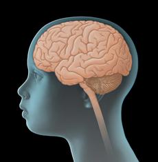
Rhabdoid tumours are rare, very aggressively growing tumours, which mostly occur in infants and toddlers during the first two years of life. They are embryonal tumours, which means that they originate from extremely immature (undifferentiated) cells. Rhabdoid tumours can develop in any tissue of the body. Most frequently (with about 65 %), however, they affect brain and spinal cord (hence the central nervous system, CNS). Apart from that, rhabdoid tumours are found, above all, in the kidneys and the liver as well as in soft tissues (such as in the neck, thigh or chestwall area).
Rhabdoid tumours of the central nervous system (CNS) are also known as “atypical teratoid rhabdoid tumours" – briefly AT/RT. According to the current classification of the World Health Organisation (WHO classification CNS5), they are classified as grade 4 tumours (out of four possible grades), thus representing especially rapidly growing malignant CNS tumours.
The following information is on rhabdoid tumours of the CNS only.
Incidence
Rhabdoid tumours of the central nervous system (AT/RT) are overall rare. In Germany, they account for about 0.6 % of all malignant diseases in children and adolescents under 18 years of age. This corresponds to an incidence rate of about 1 per million children. According to data of the European Registry for Rhabdoid Tumours (EU-RHAB), 15–22 patients per year have been newly diagnosed with AT/RT in Germany over the past years (for more information on EU-RHAB, please see chapter “Therapy optimising trials and registries”).
AT/RT appear in almost all age groups, however, infants and toddlers in their first two years of life are most frequently affected (with about 80 %). In children under one year of age, AT/RT are even the most common CNS tumours, according to data acquired by the German Childhood Cancer Registry (Mainz). From the age of six, the disease is rather rare. The average age at diagnosis is about 1.5 years. Boys are slightly less affected than girls (gender ratio: 0.9:1).
Site and spread
According to data of the EU-RHAB Registry (collected from 2005–2024), AT/RT develop, in about 50 % of cases, in the so-called cerebellopontine angle in the area of the cerebellum or the brain stem (that is, infratentorially), from where they invade into adjacent structures. About 34 % of the tumours are found above the cerebellar tentorium (supratentorially), for example within the hemispheres of the cerebrum. The remaining AT/RT distribute between the area of the pineal gland, the diencephalon, the spinal cord, or occur at multiple sites (multifocal). There are also tumours for which the exact primary site cannot be determined precisely due to their size. In older patients, supratentorial rhabdoid tumours occur more frequently than infratentorial rhabdoid tumours.
About 20–30 % of patients with AT/RT may present with metastases at diagnosis [see metastasis], predominantly within the central nervous system. Metastasis outside the CNS, on the other hand, is extremely rare and occurs almost exclusively in children with a predisposition for these tumours (a so-called rhabdoid tumour predisposition syndrome). In about 10–15% of patients with this syndrome, rhabdoid tumours also develop simultaneously in other parts of the body (multifocal, synchronous tumour involvement), which is not considered as metastasis.
Causes
The causes leading to the development of a rhabdoid tumour are not fully understood yet. A known fact, however, is that almost all (meaning over 90–95 % of) rhabdoid tumours – regardless of their localisation in the body – harbour a structural (epigenetic) alteration of a certain gene on chromosome 22. The affected gene is the SMARCB1 gene (also known as INI1), which encodes the protein SMARCB1/INI1. This protein plays a major role in controlling cellular mechanisms such as cell growth and differentiation. The genetic defect (mutation) leads to loss of protein function and subsequently to predisposition to malignant transformation and tumour development.
Most of the time, the mutated SMARCB1 gene is only found in the tumour cells following spontaneous malignant transformation of a somatic cell. Less frequently (in 25–30 % of patients), the germline (germ cells) and thus all cells in the body are affected (germline mutation). Both a spontaneous genetic change in the patient’s germline during embryonal development and (very rarely) a defect inherited from a parent are considered as causes. In both cases the disease is hereditary, meaning that the mutated gene and thus the predisposition to develop a rhabdoid tumour can be passed on to the offspring. Experts communicate this as “Rhabdoid Tumour Predisposition Syndrome" (RTPS). However, not all individuals harbouring a SMARCB1 mutation will develop a rhabdoid tumour.
Aside from SMARCB1 mutations, SMARCA4 mutations (also known as BRG1) on chromosome 19 very rarely cause the tumour disease as well. This mutation is frequently inherited from a parent. Depending on which type of mutation has been found (SMARCB1 oder SMARCA4), the syndrome is either called RTPS1 or RTPS2.
Good to know: In case of rhabdoid tumour predisposition syndrome (RTPS1 oder 2), thus a germline mutation, the patient’s siblings are probably also at an increased risk of developing the disease. For both kinds of the syndrome are inherited in an autosomal dominant manner, which means that each child of a parent with RTPS has a 50 % risk of carrying the familial germline mutation as well. Therefore, genetic councelling for the patient’s whole family is recommended when a rhabdoid tumour is suspected (see chapter “Diagnosis”).
Symptoms
Due to the uncontrolled and aggressive growth pattern of rhabdoid tumours, symptoms typically develop and deteriorate within a few weeks. The presenting symptoms of these rhaboid tumours (AT/RT) (like other tumours of the central nervous system) primarily depend on the patient’s age, the site and size of the tumour as well as its pattern of spread within the central nervous system (CNS). The following general (nonspecific) and local (specific) symptoms can occur:
General (nonspecific) symptoms
Unspecific general symptoms occur independently of the tumour’s location. They may be similar to and therefore mimic other, non-CNS diseases. Infants and toddlers, as the most frequently affected age-group, typically present with shrill cries, irritability, fatigue, lethargy, loss of appetite, vomiting, developmental delay and/or failure to thrive. Wry neck (torticollis) as a result of cranial nerve impairment is also a common symptom. Older kids may complain about headaches and/or back pain as well as dizziness. General symptoms of a child or adolescent with a CNS tumour may also include increasing fatigue, inability to concentrate, school problems, mood swings, and character changes.
Major reason for these symptoms is the slowly but continuously increasing intracranial pressure (ICP). Elevated ICP may be caused by the growing, thus more and more space-occupying tumour within the bony skull, but also by the tumour blocking the regular flow of the cerebrospinal fluid, thereby forming hydrocephalus. In babies or small children with soft spots (open fontanelles), elevated intracranial pressure and hydrocephalus typically present with a bulging fontanelle or a larger than expected head circumference (macrocephalus), respectively.
Local (specific) symptoms
Local symptoms may indicate the tumour location and, thus, which functional regions of the CNS may be affected. Therefore, a rhabdoid tumour in the cerebellum can cause impaired movement, visual deficits, dizziness and gait disturbances, whereas such a tumour in the hemispheres (cerebrum) can be associated with motor and sensory deficits (one-side paralyses, hemiplegia), seizures, speech disorders and behavioural problems, to name a few. A tumour in in the spinal cord area may cause serious back pain as well as palsies of various types (such as bladder and bowel incontinence).
Good to know: Not all patients presenting with one or more of the symptoms mentioned above do have a rhabdoid tumour or another type of brain tumour. Many of these symptoms may also occur with other, harmless diseases that are not associated with a brain tumour at all. However, if certain symptoms persist or get worse (for example repetitive headaches or rapid increase of head circumference in a young child), a doctor should be seen to find the underlying reason. In case it is a rhabdoid tumour or some other brain tumour, treatment should be started as soon as possible.
Diagnosis
If the paediatrician thinks that the young patient’s history (anamnesis) and physical examination are suspicious of a tumour of the central nervous system (CNS), the child should immediately be referred to a hospital with a childhood cancer program (paediatric oncology unit), where further diagnostics can be initiated and performed by childhood cancer professionals. Very close collaboration between various specialists (such as paediatric oncologists, paediatric neurosurgeons, paediatric radiologists, to name a few) is required, both to find out, whether the patient really suffers from a malignant CNS tumour and, if so, to determine the tumour type and the extension of the disease. Knowing these details is absolutely essential for optimal treatment and prognosis.
Imaging tests to confirm tumour existence
The initial diagnostic procedures for a young patient presenting with a suspected CNS tumour at a childhood cancer centre include another assessment of the patient’s history, a thorough physical/neurological exam and imaging diagnostics, such as ultrasound (sonography) and magnetic resonance imaging (MRI). Rapid clarification may require computed tomography (emergency CT). These tests help to find out exactly whether the patient has a tumour of the central nervous system. Also, localisation and extent of the tumor as well as its demarcation regarding adjacent tissue can be diagnosed by these imaging techniques very well.
Tissue extraction (biopsy) to secure diagnosis
Final diagnosis of a CNS tumour requieres the removal of tumour tissue (biopsy). This usually requires surgery. The obtained tissue will be analysed under the microscope (histologically) as well as with immunohistochemical, cytogenetic and molecular genetic methods. In particular, proof of SMARCB1 or SMARCA4 mutation facilitates establishing the diagnosis of a rhabdoid tumour (see chapter “Causes”). In the context of immunohistochemical analysis, loss of the SMARCB1 or SMARCA4 protein as a result of the mutation can be determined by specific staining of the tumour cells. Cytogenetic and molecular genetic testing provides direct proof of the genetic defect.
Once a SMARCB1 or SMARCA4 mutation has been confirmed for the tumour tissue, blood cells (or any other tumour-free tissue) will be tested for the mutation as well in order to rule out a germline mutation and rhabdoid tumour predisposition syndrome (RTPS), since in case of a germline mutation, the blood cells (or other tissue cells) do contain the mutation as well. The germline mutation is particularly suspected in children under two years of age, in patients with tumours at multiple sites (synchrone tumours) or in case of a family history of tumour diseases, respectively.
In case a family history of rhabdoid tumour predisposition syndrome (RTPS1 or RTPS2) is known prior to biopsy, testing only the patient’s blood for mutation can be sufficient to confirm diagnosis, since a malignant tumour revealed by diagnostic imaging and with SMARCB1 or SMARCA4 mutation in the patient’s blood cells is most likely an AT/RT. However, blood tests cannot replace biopsy and may, therefore, be an option but in very rare situations (for example, if the child is to ill to be operated).
Aside from the examination of tumour tissue for a SMARCB1 or SMARCA4 mutation, the determination of the DNA methylation profile is becoming increasingly important. With the help of this molecular genetic method, AT/RT can be divided into three different molecular (epigenetic) subtypes (ATRT-MYC, ATRT-SHH, ATRT-TYR), some of which also behave differently regarding their growth pattern (see chapter "Therapy planning").
Important to know: diagnosis of a germline mutation not only affects a patient’s prognosis, but maybe also other family members, especially regarding the need for subsequent screening tests (see recommendations in case of a rhabdoid tumour predisposition syndrome below).
Tests to assess spread of disease (tumour staging)
Once the diagnosis of a rhabdoid tumour has been confirmed, additional tests are required to assess the extent of the disease within body (stage of disease), Since rhabdoid tumours are known to metastasize frequently and also may be found at multiple sites already at diagnosis, for example in the central nervous system (CNS) and simultaneously in the kidneys (synchronous rhabdoid tumour), the initial finding of a single tumour is always followed by diagnostic imaging of the whole body.
Usually, a magnetic resonance imaging (MRI) of the complete CNS (brain and spine) is performed to rule out metastasis within the CNS, furthermore, at least once, a total body scan to assess macroscopic distant metastases and/or synchronous rhabdoid tumours. Apart from that, mostly after surgery, the cerebrospinal fluid (CSF) is microscopically checked for tumour cells in the spinal cord (which are not visible by MRI scan). Cerebrospinal fluid is mostly obtained from the spine in the lower back (lumbar puncture), since the risk of the puncture needle damaging the spinal cord is lowest at the lower back level.
Tests before treatment begins
In preparation for the intensive treatment of the brain tumour, further investigations are performed, such as electrocardiography (ECG) and/orechocardiography to check cardio function or electroencephalography (EEG) to test brain function. Furthermore, blood tests are needed to assess the patient’s general health condition and to check whether the function of certain organs (such as liver and kidneys) is affected by the disease and whether there are any metabolic disorders to be considered prior or during therapy. Any changes occurring during the course of treatment can be assessed and managed better based on the results of those initial tests, which thus help to keep the risk of certain treatment-related side effects as low as possible.
If the child's general condition is stable, neuropsychological examinations (for example by means of questionnaires) can also be carried out at the time of diagnosis [see neuropsychology]. These serve to record the child's level of development and cognitive abilities as well as any abnormalities as a basis for future surveys. Examinations on quality of life may also be included.
Good to know: Not every patient needs the complete check-up. On the other hand, tests might be added that haven't been mentioned here, depending on the individual situation of the patient. Your caregivers will inform you and your child, which diagnostic procedures are individually required in your case and why.
Recommendations if a rhabdoid tumour predisposition syndrome is suspected
If a patient is diagnosed with a SMARCB1 germline mutation – and, subsequently, is at an increased risk for developing a rhabdoid tumour (rhabdoid tumour predisposition syndrom, RTPS) –, one possible reason may be that the disease has been inherited by one parent. In most of the cases, however, it is more likely that the genetic defect is a result of a new SMARCB1 mutation.
If inheritance is the case, both the patient’s parents and siblings are at a higher risk to develop a rhabdoid tumour as well (see chapter „Causes“). In order to rule out this risk or to diagnose it as early as possible, certain blood tests will be recommended to both parents first. If those come back positive for a germline mutation in one of the parents, the patient’s siblings should also be tested. These tests are usually done in human genetics laboratories. Also, genetic councelling is recommended.
If a rhabdoid tumour predesposition syndrome is known in the family and a germline mutation is found in a so far healthy family member, it is recommended that the affected child(ren) is (are) closely monitored from birth by regular physical/neurological examinations, magnetic resonance imaging and ultrasound (of the head, abdomen and chest).
Treatment planning
After the diagnosis has been confirmed, therapy is planned. In order to design a highly individual, risk-adapted treatment regimen for the patient, certain individual factors influencing the patient’s prognosis (called risk factors or prognostic factors) are being considered during treatment planning (risk-adapted treatment strategy).
One important prognostic factor is the patient‘s age at diagnosis: it determines treatment intensity and, therefore, has an impact on the patient’s chances of survival. For example, radiotherapy – a very efficient treatment method for rhabdoid tumours – is only a limited option for children under the age of three and no option at all for children under the age of 12 months. Also, the tolerance of this treatment is significantly reduced in very young children when compared to other methods (such as surgery, chemotherapy).
Another important prognostic factor is the molecular subtype of AT/RT, since this may also impact the course of the disease (see chapter "Diagnosis / Tissue extraction"). For example, patients with the ATRT-TYR subtype usually have a better chance of cure than patients with one of the other two subtypes (ATRT-MYC, ATRT-SHH). In summary, knowledge of the subtype allows drawing conclusions on prognosis.
Localization, size and spread of the tumour as well as its type (hereditary or non-hereditary) are further factors affecting prognosis. Hereditary disease (that is, a rhabdoid tumour predisposition syndrome, RTPS), is known as an unfavourable prognostic factor and so is metastasis at the timepoint of tumour diagnosis. Both scenarios are associated with a lower probability of eliminating all tumour manifestations (which is known to promote favourable prognosis). Response of the disease to chemotherapy is also an important prognostic factor.
All these factors are included in treatment planning in order to achieve the best outcome possible for each patient.
Treatment
Treatment of children and adolescents with rhabdoid tumour should take place in a children's hospital with a paediatric oncology program. Only such a childhood cancer centre provides highly experienced and qualified staff (doctors, nurses and many more), since they are specialised and focussed on the diagnostics and treatment of children and teenagers with cancer according to the most advanced treatment concepts. The doctors in these centres collaborate closely with each other. Together, they treat their patients according to treatment plans (protocols) that are continuously optimised.
The goal of the treatment is to achieve high cure rates while avoiding side effects as much as possible. Considering the usually very young age of patients with a rhabdoid tumour, this is a huge challenge. Infants and toddlers are highly vulnerable; they suffer severely of acute side effects and long-term sequelae of the aggressive therapy and are therefore hard to treat. Hence, the most important step prior to or during therapy is to decide whether treatment should be initiated or continued, respectively, and if so, whether the goal is cure (curative approach) or managing symptoms (palliative therapy).
From 2007 until the opening of the international therapy trial SIOPE ATRT01 in 2021, patients with rhabdoid tumour received treatment according to a standardized therapy protocol under the roof of the EU-RHAB Registry (so-called consensus treatment strategy, see chapter "Therapy optimising trials and registries“). The following information on therapy is based on this consensus treatment strategy and the current guidelines of the Society of Paediatric Oncology and Haematology (GPOH) for AT/RT.
Treatment methods
Treatment options for patients with rhabdoid tumour include surgery, chemotherapy and radiotherapy. For some patients, high-dose chemotherapy followed by autologous stem cell transplantion may be an option, too. The individual treatment choice is based on the patient’s age and general health status as well as on the tumour type and its extent at diagnosis and after surgery.
Neurosurgery and radiotherapy are the most promising therapy methods and of highest impact in the treatment of patients with rhabdoid tumour. Yet, surgery or radiation cannot be performed in every child. Radiotherapy, for example, is only possible from a certain age. Chemotherapy (and, for some patients, high-dose chemotherapy) can help improve prognosis and – particularly in very young children – delay or even completely avoid radiotherapy.
Surgery
First step when treating a patient with rhabdoid tumour is maximal neurosurgical tumour removal [see neurosurgery], if possible, since the extent of tumour resection seem to have a major impact on the subsequent course of the disease. The more radically the tumour can be resected, the higher are the chances of long-term survival. If the tumour is localized and has not spread, second look surgery to achieve gross total removal is an option. Unfortunately though, complete resection is impossible in most cases (in about 70 % of patients) without also removing healthy tissue and without jeopardizing the patient’s quality of life. This is related to the frequently unfavourable tumour location, the patient’s young age as well as the frequent presence of metastases [see metastasis] at the timepoint of tumour diagnosis.
Aside from tumour removal, neurosurgery also includes the implantation of a catheter into one of the brain’s ventricles (cerebral ventricles) for subsequent chemotherapy. The catheter is connected to a small, dome-shaped, soft plastic device (Ommaya reservoir or Rickham reservoir) placed under the patient’s scalp, through which chemotherapy can directly be delivered into the cerebrospinal fluid (see next chapter).
Chemotherapy
Surgery is followed by intensive chemotherapy in order to improve the patient’s chances of cure. Chemotherapy uses drugs (so-called cytostatic agents or cytostatics) that can kill fast-dividing cells, such as cancer cells, or inhibit their growth, respectively. In order to optimize treatment efficacy, combinations of different cytostatics are given in different treatment blocks.
Standard therapy according to the current guideline consists of two phases, an induction and a consolidation phase, and may include up to 12 treatment blocks. Induction therapy aims at a maximum possible control of the tumour and/or the tumour cell count, while the aim of consolidation therapy is to maintain (consolidate) the results gained by induction. The therapy includes the agents doxorubicine (DOX) and combinations of ifosfamide, carboplatin and etoposide (briefly: ICE) or vincristine, cyclophosphamide and actinomycin D (briefly: VCA), respectively, which are given intravenously in alternation.
By the intravenous route, they get distributed in the blood system and can, thus, eliminate tumour cells throughout the whole body (systemic chemotherapy). In addition to this approach, some chemotherapy (methotrexate, MTX) is given directly into the cerebrospinal fluid, which surrounds both brain and spinal cord. This intraventricular or intrathecal chemotherapy is necessary, because most chemotherapeutic agents cannot pass the barrier between blood and brain (blood-brain barrier).
Radiotherapy
Depending on the patient’s age at the timepoint of treatment, radiotherapy may be recommended during or after chemotherapy. Radiotherapy is carried out using energy-rich, electromagnetic radiation, given through the skin to the tumour region. Radiation causes DNA damage in tumour cells, thereby leading to cell death.
Aside from complete surgical tumour removal, radiotherapy is one of the most important and successful measures for treatment of a rhabdoid tumour. Studies have shown that patients benefit from early radiation. However, its application is limited due to treatment-induced late effects. This particularly affects infants and toddlers, whose brains are still developing: radiation early in life results in serious impairment of normal cognitive development. Therefore, chemotherapy and, if applicable, even high-dose chemotherapy are used to delay radiotherapy as long as possible.
If radiotherapy is an option, timing, volume and type (photons versus protons) are determined based on the patient’s age, the vulnerability of the tissue and prognostic factors.
According to the current guidelines for AT/RT, radiotherapy is considered for children 12–18 months of age with localized disease. These patients will receive a total dose of 54 gray (Gy) to the tumour region. For children whose tumour has spread within the central nervous system via the cerebrospinal fluid (leptomeningeally metastasized tumours), radiation of the brain and spinal cord (craniospinal radiotherapy) are considered once they are three years old. Fore these patients, a dose of 35.2 Gy to the whole central nervous system plus an additional dose (boost) to the primary tumour site (up to a total dose of 55 Gy) are recommended.
Modern radiation techniques, such as intensity-modulated radiotherapy (IMRT), help minimise the damage of healthy tissue. For some patients, radiotherapy with protons instead of conventional radiotherapy (with photons) may be an option, for example for very young children or in case proton therapy is expected to have a clear advantage compared to conventional radiotherapy. This type of radiotherapy allows to reduce the effects of radiation in healthy tissue even better and is, therefore, gaining an increasing importance in the treatment of children and teenagers with solid tumours.
Note on the SIOPE ATRT01 trial: This study examines particularly, whether radiation-induced long-term sequelae in young children can be avoided by the use of high-dose chemotherapy only (no radiotherapy) without jeopardizing the patient's chances of cure. Hence, the feasibility of high-dose chemotherapy only versus the current regimen of combined chemo- and radiotherapy (12 courses of chemotherapy plus radiotherapy) will be analysed. The study will include children aged 12–35 months who have no metastases and no rhabdoid tumour predisposition syndrome (RTPS).
High-dose chemotherapy and autologous stem cell transplantation
For some patients, high-dose chemotherapy as a consolidation treatment followed by autologous stem cell transplantation may be an option instead of the conventional chemotherapy described above. In this case, the patient receives treatment with carboplatin and thiotepa (CARBO/TT) after six cycles of standard chemotherapy (induction therapy, see chapter “chemotherapy”). The chemotherapy doses of this regimen are considered high enough to also eliminate otherwise treatment-resistant tumour cells in the body.
Since the high doses of cytostatics given according to this treatment strategy also lead to the destruction of the blood-forming cells in the bone marrow, the patient will receive blood-forming stem cells in a second step. These blood stem cells are obtained from the patient’s blood or bone marrow prior to high-dose chemotherapy and are given back right after this treatment (so-called autologous stem cell transplantation, SCT).
Precondition for carrying out this treatment is, however, that most of the tumour load has been destroyed by the preceding standard regimen, thereby having achieved a so-called remission. Also, the patient’s age and general clinical condition are of relevance, for the treatment will be stressful and associated with risks.
It is still unclear so far whether the high-dose regimen provides a prognostic benefit over the standard chemotherapy (in children under 12 months of age) or the conventional chemo-/radiotherapy (in children older than 12 months). This question is currently being addressed by the European Umbrella trial SIOPE ATRT01, which has been open since 01/06/2021.
Note on the SIOPE ATRT01 trial: The study will examine whether high-dose chemotherapy (without radiation) for consolidation achieves the same results as combined radiotherapy/chemotherapy in children aged 12–35 months – following induction chemotherapy. For children under 12 months of age or children who are not eligible for radiotherapy, the study will examine whether high-dose chemotherapy leads to better survival rates than continuation of conventional chemotherapy in the consolidation.
For further, general information on stem cell transplantation, please see here.
New treatment approaches
Despite the currently available intensive treatment modalities, cure rates for children with AT/RT are unsatisfying. This particularly applies to high-risk patients (young age at diagnosis, unfavourable molecular subtype, germline mutation and/or metastasised tumour). In addition, the intensive treatment does not only cause acute side effects, but also long-term sequelae (such as hormonal deficits, which are associated with certain developmental delays, or impairments in hearing or vision). This may seriously impair the patients’ quality of life.
Scientists keep studying these tumours intensely to find new agents and treatment modalities. The current research focuses on the molecular mechanisms leading to the developments and growth of rhabdoid tumours. The analysis of cellular signalling pathways that are altered in rhabdoid tumours have helped identifying different agents, which may be of benefit in the treatment of rhabdoid tumours. Promising new treatment strategies will be examined in the framework of clinical studies.
For patients with a relapse of an AT/RT, various approaches apply to individual treatment attempts. These include epigenetically active agents like decitabine on the one hand, but also the drug ribociclib and so-called checkpoint inhibitor. Another therapy option is a metronomic therapys, for example within the framework of the MEMMAT protocol.
Unfortunately, there are currently only few phase I/II studies open for children. The NivEnt study, for example, only includes children with a certain molecular AT/RT subtype (ATRT-MYC). The researchers at the EU-RHAB Registry are working hard to find new drugs and get them approved for clinical use. This should always be carried out as clinical studies (see also next chapters).
Therapy optimising trials and registries
In the large paediatric treatment centres, children and teenagers with rhabdoid tumour receive therapy according to guidelines and/or standardized treatment plans (protocols). These protocols are designed by experts with the goal to improve the patient’s prognosis and are usually applied within therapy optimising trials or registries. Therapy optimising trials are standardised and controlled clinical trials that aim at steadily developing and improving treatment concepts for sick patients based on the current scientific knowledge.
Patients who cannot participate in any study, for example because none is available or open for them at that time, or since they do not meet the required inclusion criteria, respectively, are often included in a so-called registry. Such a registry primarily serves to acquire all clinical, molecular genetic and treatment-associated patient data in ordert o gain a better understanding oft he tumour biology. Furthermore, the registry center supports the doctors at site with (non-commital) treatment recommendations based on the most recent data on best treatment options, in order to provide the patient with optimal therapy even without the framework of a clinical study.
Currently available for patients with AT/RT are the trial SIOPE ATRT 01 and the EU-RHAB registry. Further trials are being developed. For more information on trials and registry please see below.
European Rhabdoid Registry (EU-RHAB Registry)
Since rhabdoid tumours are very rare, experts of the Society of Paediatric Oncology and Haematology (GPOH) initiated during a consensus conference in Italy in 2007 that all patients with rhabdoid tumours in a part of Europe should be registered with the European Rhabdoid Registry (EU-RHAB Registry) and treated according to a standardized strategy (consensus strategy). The treatment regimen was designed by an international competence network and is outlined in the chapter “Treatment” as well as in the current guideline for AT/RT. It has been validated as standard therapy, unless the patient is not treated within the experimental arm of a trial, and requires adjustment to the individual patient.
Numerous countries worldwide are currently registering their patients with this EU-RHAB Registry. Treatment centres in Germany, where children and adolescents with cancer are being treated, are legally obliged to register their patients with the appropriate studies or registries. The European Registry headquarters are located at the University Hospital in Augsburg, Germany, and the principal investigator is Prof. Dr. Dr. med. Michael C. Frühwald. Please note: Patient recruitment has been stopped on a temporary basis since 24/04/2023! The new registry (EU-RHAB 2.0) is currently being evaluated by the Ethics Committee.
Therapy trial SIOPE ATRT01
In July 2021, an international, multicentric trial – developed in the framework of the EU-RHAB registry – has been opened for children with AT/RT: the umbrella study SIOPE ATRT01. In this context, the term "umbrella" means that the study consists of several parts and assigns the affected patients to a therapy arm depending on their individual risk profile.
The umbrella study enrolls all European patients aged 0–17 years. The integrated randomised therapy optimising trial is intended for patients aged 12–35 months [see randomisation]. Numerous treatment centres in Germany and in almost all other European countries (including France, Switzerland, England, Spain and Portugal) are participating in the study. The study centre is located at the University Hospital in Augsburg, Germany (principal investigator: Prof. Dr. Dr. med. Michael C. Frühwald).
New trials
Within the framework of EU-RHAB, new studies are also being developed based on previously acquired data. A Phase I/II study is currently under development to test a new substance that is presumably effective in rhabdoid tumours. In addition, a new treatment protocol is being developed for the treatment of patients with disease relapse.
Prognosis
The chances of cure (prognosis) for children with a newly diagnosed rhabdoid tumour of the central nervous system have significantly improved thanks to standardised, multimodal treatment concepts within the framework of EU-RHAB. Patients treated in the most favourable risk (therapy) group meanwhile present with 5-year-survival rates of more than 71.5 %. For patients with unfavourable prognostic factors, however, prognosis is still not favourable, despite the intensive therapy. According to data of the EU-Rhab Registry, the average overall 5-year-survival rate (for all risk groups taken together) is about 35–40 %.
Important prognostic factors are the patient’s age at diagnosis, tumour type (hereditary or non-hereditary, molecular subtype) as well as tumour size, site and extent and, thus, possibility of complete tumour removal (see chapter “Treatment planning”). Survival rates are thus different in individual patients:
- Patients with localized, non-metastasised, surgically removable and non-hereditary rhabdoid tumour, who are older than three years of age at diagnosis, usually have favourable probabilities of cure, given that the tumour can be completely removed and early radiotherapy is possible.
- Children between their first and third birthday with the same co-factors have a comparably higher risk of relapse and thus less favourable outcomes.
- Prognosis is particularly unfavourable for infants and toddlers under one year of age, especially if they present with a certain molecular AT/RT subtype.
- The same applies to all other patients with unfavourable prognostic factors, that is, a high-risk rhabdoid tumour. These include patients with germline mutation, and thus a predisposition for the development of rhabdoid tumour, as well as patients with surgically unremovable primary tumour or metastasised disease, respectively.
However, patients who do benefit from treatment (surgery, chemotherapy, in some cases high-dose chemotherapy and radiotherapy) despite of unfavourable prognostic factors, so that long-term survival may be possible. New molecular treatment approaches are currently being analysed in the frame-work of therapy optimising trials. The goal is to optimize cure rates also for high-risk patients.
Note: The survival rates mentioned in the text above are statistical values. Therefore, they only provide information on the total cohort of patients with this kind of brain tumours. They do not predict individual outcomes.
 PDF: Information on rhabdoid tumours of the central nervous system (AT/RT) (344KB)
References 
- Ronckers CM, Spix C, Grabow D, Erdmann F: German Childhood Cancer Registry - Annual Report 2022 (1980-2021). Institute of Medical Biostatistics, Epidemiology and Informatics (IMBEI) at the University Medical Center of the Johannes Gutenberg University Mainz 2025 [URI: https://www.kinderkrebsregister.de/ fileadmin/ kliniken/ dkkr/ pdf/ jb/ jb2022/ JB_2022_final.pdf]
- Traunwieser T, Loos E, Ottensmeier H, Gastberger K, Nemes K, Mynarek M, Bison B, Kandels D, Neumayer P, Neumann-Holbeck A, Lüttich P, Baust K, Faulstich-Ritter K, John R, Kreisch A, Landmann J, Manteufel E, Nest A, Prüfe J, Schubert L, Stamm W, Timmermann B, Gerss J, Rutkowski S, Schlegel PG, Eyrich M, Gnekow AK, Frühwald MC: Survivors of infant atypical teratoid/rhabdoid tumors present with severely impaired cognitive functions especially for fluid intelligence and visual processing: data from the German brain tumor studies. Pediatric blood & cancer 2024, 71:e30910 [PMID: 38342954]
- Frühwald MC et al.: S1-Leitlinie „Atypische Teratoide / Rhabdoide Tumoren“ der Gesellschaft für Pädiatrische Onkologie und Hämatologie. Registernummer 025-037 AWMF online 2024 [URI: https://register.awmf.org/ assets/ guidelines/ 025-037l_S1_Atypische-Teratoide-Rhabdoide-Tumoren_2024-05.pdf]
- Rutkowski S, Pfister S, Frühwald M, Fleischhack G, Korinthenberg R, Bison B, Hahn G, Mentzel H-J, Langen K-J, Hernáiz-Driever P, Pietsch T: Leitsymptome und Diagnostik der ZNS-Tumoren im Kindes- und Jugendalter. Gemeinsame Leitlinie der Gesellschaft für Neuropädiatrie und der Gesellschaft für Pädiatrische Onkologie und Hämatologie AWMF online, 2024 [URI: https://register.awmf.org/ assets/ guidelines/ 025-022l_S1_Leitsymptome-Diagnostik-ZNS-Tumoren-Kinder-Jugendliche_2024-06.pdf]
- Louis DN, Perry A, Wesseling P, Brat DJ, Cree IA, Figarella-Branger D, Hawkins C, Ng HK, Pfister SM, Reifenberger G, Soffietti R, von Deimling A, Ellison DW: The 2021 WHO Classification of Tumors of the Central Nervous System: a summary. Neuro-oncology 2021 Aug 2; 23: 1231 [PMID: 34185076]
- Frühwald MC, Nemes K, Boztug H, Cornips MCA, Evans DG, Farah R, Glentis S, Jorgensen M, Katsibardi K, Hirsch S, Jahnukainen K, Kventsel I, Kerl K, Kratz CP, Pajtler KW, Kordes U, Ridola V, Stutz E, Bourdeaut F: Current recommendations for clinical surveillance and genetic testing in rhabdoid tumor predisposition: a report from the SIOPE Host Genome Working Group. Familial cancer 2021,online ahead of print [PMID: 33532948]
- Frühwald MC, Hasselblatt M, Nemes K, Bens S, Steinbügl M, Johann PD, Kerl K, Hauser P, Quiroga E, Solano-Paez P, Biassoni V, Gil-da-Costa MJ, Perek-Polnik M, van de Wetering M, Sumerauer D, Pears J, Stabell N, Holm S, Hengartner H, Gerber NU, Grotzer M, Boos J, Ebinger M, Tippelt S, Paulus W, Furtwängler R, Hernáiz-Driever P, Reinhard H, Rutkowski S, Schlegel PG, Schmid I, Kortmann RD, Timmermann B, Warmuth-Metz M, Kordes U, Gerss J, Nysom K, Schneppenheim R, Siebert R, Kool M, Graf N: Age and DNA-methylation subgroup as potential independent risk factors for treatment stratification in children with Atypical Teratoid/Rhabdoid Tumors (ATRT). Neuro-oncology 2020, [PMID: 31883020]
- Kuhlen M, Wieczorek D, Siebert R, Frühwald MC: How I approach hereditary cancer predisposition in a child with cancer. Pediatric blood & cancer 2019, 66:e27916 [PMID: 31342632]
- Frühwald MC, Furtwängler R: Das Europäische Rhabdoidregister – Basis für klinischen Fortschritt in der Behandlung einer sehr seltenen Tumorerkrankung. WIR - die Zeitschrift der Deutschen Leukämie-Forschungshilfe e.V. und der Deutschen Kinderkrebsstiftung 2018, 2/18
- Nemes K, Clément N, Kachanov D, Bens S, Hasselblatt M, Timmermann B, Schneppenheim R, Gerss J, Siebert R, Furtwängler R, Bourdeaut F, Frühwald MC, EU-RHAB consortium: The extraordinary challenge of treating patients with congenital rhabdoid tumors-a collaborative European effort. Pediatric blood & cancer 2018, 65:e26999 [PMID: 29418059]
- Nemes K, Frühwald MC: Emerging therapeutic targets for the treatment of malignant rhabdoid tumors. Expert opinion on therapeutic targets 2018, 22: 365 [PMID: 29528755]
- Frühwald MC, Hasselblatt M: Rhabdoide Tumoren des ZNS, der Nieren und des Weichteilgewebes. in: Niemeyer C, Eggert A (Hrsg.): Pädiatrische Hämatologie und Onkologie, Springer-Verlag GmbH Deutschland 2. vollständig überarbeitete Auflage 2018, 402 [ISBN: 978-3-662-43685-1]
- Nemes K, Bens S, Bourdeaut F, Hasselblatt M, Kool M, Johann P, Kordes U, Schneppenheim R, Siebert R, Frühwald MC, In: Adam MP, Ardinger HH, Pagon RA, Wallace SE, Bean LJH, Stephens K, Amemiya A (eds): Rhabdoid Tumor Predisposition Syndrome. GeneReviews 2017 [PMID: 29215836]
- Bartelheim K, Nemes K, Seeringer A, Kerl K, Buechner J, Boos J, Graf N, Dürken M, Gerss J, Hasselblatt M, Kortmann RD, Teichert von Luettichau I, Nagel I, Nygaard R, Oyen F, Quiroga E, Schlegel PG, Schmid I, Schneppenheim R, Siebert R, Solano-Paez P, Timmermann B, Warmuth-Metz M, Frühwald MC: Improved 6-year overall survival in AT/RT - results of the registry study Rhabdoid 2007. Cancer medicine 2016, [PMID: 27228363]
- Frühwald MC, Biegel JA, Bourdeaut F, Roberts CW, Chi SN: Atypical teratoid/rhabdoid tumors-current concepts, advances in biology, and potential future therapies. Neuro-oncology 2016, 18: 764 [PMID: 26755072]
- Benesch M, Bartelheim K, Fleischhack G, Gruhn B, Schlegel PG, Witt O, Stachel KD, Hauch H, Urban C, Quehenberger F, Massimino M, Pietsch T, Hasselblatt M, Giangaspero F, Kordes U, Schneppenheim R, Hauser P, Klingebiel T, Frühwald MC: High-dose chemotherapy (HDCT) with auto-SCT in children with atypical teratoid/rhabdoid tumors (AT/RT): a report from the European Rhabdoid Registry (EU-RHAB). Bone marrow transplantation 2014, 49: 370 [PMID: 24419520]