X-Ray
Author: Gesche Tallen, MD, PhD, Editor: Maria Yiallouros, Reviewer: Prof. Dr. med. Dr. h. c. Günter Henze, English Translation: Hannah McRae, Last modification: 2015/04/22 https://kinderkrebsinfo.de/doi/e74284
X-ray techniques have been used in cancer diagnostics for a long time. Despite of continuous progress in the development of other imaging procedures, they still serve as a standard diagnostic method in paediatric oncology.
X-ray techniques use a form of radiation with high energy that is called electro-magnetic radiation. While penetrating the patient's body, x-rays are being absorbed by the different organs. The level of this absorption is dependent on the densitiy of each type of tissue. Those x-rays that passed a certain body region are caught on a special photographic plate (x-ray film) in order to detect them and provide a picture of the organ of interest.
There are three major levels of tissue density, that can be seen on an X-ray film since they show different monochrome (black and white) shades:
- density of bone (such as ribs, skull): white
- density of soft tissue (such as the heart): different nuances of grey
- density of air (for example within the lungs or intestines): black
In a child or teenager with cancer, X-ray techniques are used to reveal abnormali-ties in tissue densities that can be indicative of cancer, cancer spread (metastasis), recurrent disease, or infections and other side-effects of the treatment.
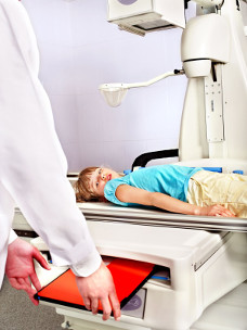
Getting an X-ray done is fast and does not hurt. However, X-rays can have negative side effects on most body tissues and cells, especially on tissues that are still growing, as they are in children and teenagers. Therefore, paediatricians have learned to make sure, that their patients get as least X-rays done as necessary.