Angiography
Author: Gesche Tallen, MD, PhD, Editor: Maria Yiallouros, Reviewer: Prof. Dr. med. Ursula Creutzig, English Translation: Hannah McRae, Last modification: 2015/01/13 https://kinderkrebsinfo.de/doi/e2036
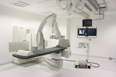
The term “angiography” refers to the visualisation of blood vessels. Techniques to visualise veins only are referred to as phlebography. To obtain images of both veins and arteries in a particular, mostly bony or dense soft tissue, region of the body a digital subtraction angiography (DSA) is performed.
Angiographic images are produced using contrast medium, which the patient receives through a catheter placed in the artery in the groin.
(© Alterfalter - Fotolia.com)
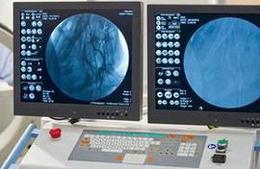
The doctors monitor the contrast medium traveling through the patient's bloodstream by looking at life X-ray images (please see X-ray) on a big monitor during the examination. This monitoring helps the radiologist to immediately detect any abnormalities in blood flow, such as clogged blood vessels (thrombosis), vascular malformations as well as vascularised tumours and the blood vessels within tumours.
The use of angiographic methods and procedures is not part of the routine diagnostics for children and adolescents with cancer. Angiography is rather used to answer special diagnostic questions, such as prior to surgical removal of a tumour that has a lot of blood vessels. Detailed information on a tumour's blood flow helps the surgeon to prevent vascular injury and bleeding.
The results of an angiography are usually analysed by a paediatric radiologist. An angiography is generally only performed on children or teenagers (under 18 years of age) after an informed consent has been obtained by the parent or legal guardian. Also, in order to obtain valid images, it requires the patient to not move during the procedure at all. Hence, most young patients are usually sedated during the procedure, so they sleep, and thus don't experience pain during the injection and don't move.
After an angiography, the patient is supposed to stay in bed for about a day keeping a special dressing in the groin in order to allow the injection to close up and prevent it from bleeding.
Use in therapy
This method can be an alternative to surgery for patients with highly vascularised tumours, such as hemangiomas and hemangioendotheliomas.
In some patients, angiographic methods can also be used to reduce a tumour's blood flow. To achieve this, the doctors use angiographic techniques (please see above) to purposely clogg up the blood vessels in the tumour (angiographic embolisation).
The efficacy of intra-arterial administration of chemotherapy using angiographic techniques in the treatment of various solid tumours (such as soft-tissue sarcomas and osteosarcomas) in children and teenagers with cancer is still being investigated.